
Welcome to the PMV Hub — a friendly corner of the internet for pet owners who want honest, easy-to-read info without the overwhelm. This is where we post blogs, pet health tips, and real talk about life with cats and dogs. Have a browse at your own pace, and if there’s a topic you’d like us to cover, just let us know via the form at the bottom. We’re always happy to take requests

Gingival Hyperplasia in Dogs and Cats: When Gum Tissue Grows Too Much

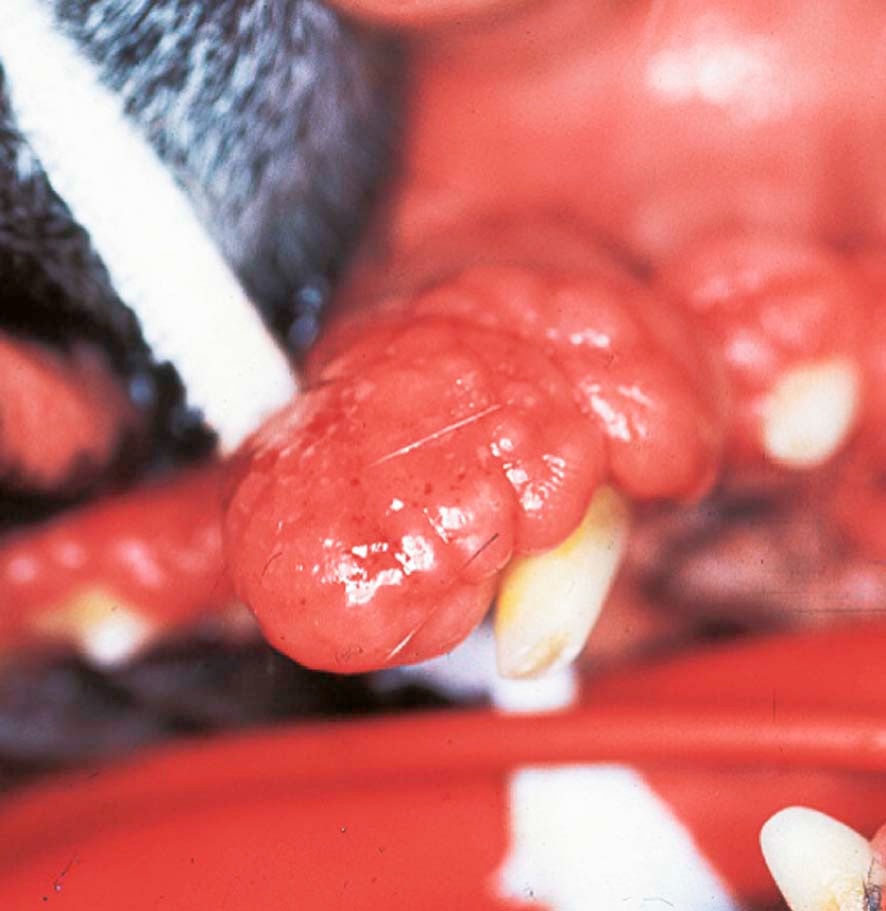
Close-up of a dog's teeth showing thickened, overgrown gum tissue characteristic of gingival hyperplasia
Gingival hyperplasia causes the gum tissue to thicken and grow upward over the tooth surfaces, creating pockets that trap bacteria and debris.
Gingival hyperplasia is a condition where a dog or cat’s gum tissue grows thicker and taller than it should, creating pockets that trap bacteria and debris. This article explains what causes it, which breeds are most affected, why it matters, and what treatment and home care look like.

Your pet’s mouth is one of the areas that most owners look at least often, and most vets wish they looked at more. Dental disease is among the most common health problems seen in dogs and cats, and one condition that sits in the “easy to miss, harder to treat once advanced” category is gingival hyperplasia, which is a fancy way of saying the gum tissue has overgrown.

This article covers what gingival hyperplasia actually is, what causes it, which animals are most at risk, what it can be confused with, how it is diagnosed and treated, and what home care looks like long-term. If you have a Boxer, or any dog or cat with gums that seem unusually thick or lumpy, it is worth reading through.

What Is Gingival Hyperplasia?

The gums are called the gingiva. They are supposed to sit snugly at the base of each tooth at a consistent, predictable height. In gingival hyperplasia, the tissue making up the gums grows beyond its normal boundaries, causing the gum margin to thicken and creep upward over the tooth surface. The word hyperplasia itself means an abnormal increase in the number of cells in a tissue, where the cells are still normal but there are simply too many of them (Niemiec et al., 2020; Zachary, 2022).

This is not a tumour. It is not cancer. It is an overgrowth of tissue types that are otherwise entirely normal, just present in excess. The overgrowth affects the fibrous tissue beneath the gum surface and the surface layer above it, so the gums become bulkier, firmer, and in some cases dramatically enlarged (Niemiec et al., 2020).

The reason it matters is what happens secondary to that overgrowth. More on that shortly.

What Causes It?

There are four main pathways that lead to gingival hyperplasia (Niemiec et al., 2020; Gorrel et al., 2013):

1.Genetics

Some dogs carry a hereditary tendency to develop this condition regardless of how clean their teeth are kept. The gum tissue responds to normal, everyday signals by growing more than it should. This is not something that can be prevented through hygiene alone. Breed predisposition is one of the strongest risk factors known, and it cannot be bred out of an individual dog.

2.Chronic Inflammation from Plaque

When plaque, which is the sticky bacterial film that constantly forms on tooth surfaces, is not removed regularly, it triggers a slow, ongoing inflammatory response in the surrounding gum tissue. Over months and years, this chronic low-grade inflammation can drive the gum tissue to expand and thicken. The overgrowth is the gum’s prolonged reaction to bacterial load, and it tends to worsen as the bacteria accumulate further in the increasingly deep crevices that the thickened gums create (Niemiec et al., 2020).

3.Medication Side Effects

Certain medications, when used long-term, alter how gum tissue cells behave, leading to overgrowth. The drug classes most commonly associated with this side effect are:

Calcineurin inhibitors, such as cyclosporine, which is used to manage immune-mediated conditions including skin disease and some eye conditions

Calcium channel blockers, such as amlodipine and nifedipine, used in the management of heart disease and hypertension, particularly in cats

Anticonvulsants, such as phenobarbital, used to manage epilepsy and seizures

If your pet is on any of these medications and you notice changes to the gum tissue, it is worth raising with your vet at the next visit (Niemiec et al., 2020; Bonagura & Twedt, 2014; Diesel & Moriello, 2015).

4.Chronic Mechanical Trauma

Repeated physical irritation to the gum tissue from abnormal tooth contacts or chewing habits can occasionally drive a localised fibrous overgrowth at a specific site. This is a less common pathway than the others (Niemiec et al., 2020).

Which Dogs and Cats Are Most Commonly Affected?

Dogs

Gingival hyperplasia is predominantly a condition of adult to senior dogs, though age of onset varies with cause (Niemiec et al., 2020; Tilley et al., 2021).

Boxers have the highest documented incidence of any breed. Approximately 30 per cent of Boxers older than five years develop hyperplastic gum lesions, making it a near-routine finding in older dogs of this breed. If you own a Boxer and are not already in the habit of looking in their mouth regularly, now is a good time to start (Niemiec et al., 2020).

Other breeds with a well-recognised predisposition include Great Danes, Collies, Doberman Pinschers, Dalmatians, and Shetland Sheepdogs. In many of these breeds, the hyperplasia tends to be generalised, meaning it affects multiple areas of the mouth rather than one isolated spot (Niemiec et al., 2020; Tilley et al., 2021).

Brachycephalic breeds, a term for flat-faced dogs such as Bulldogs, Pugs, and French Bulldogs, are also associated with higher rates of notable oral soft tissue changes. The crowding and misalignment that come with a compressed facial structure create additional opportunities for abnormal gum responses (Niemiec et al., 2020; Ryu et al., 2024).

Cats

Generalised gingival hyperplasia is uncommon in adult cats. When it does occur in cats, it most frequently affects young cats around the age of six to eight months, during or shortly after the eruption of permanent teeth. This is often referred to as feline juvenile hyperplastic gingivitis. Breeds seen in this group include Maine Coons, Abyssinians, Tonkinese, and domestic shorthairs (Niemiec et al., 2020; Soltero-Rivera et al., 2023; Peterson & Kutzler, 2011).

What Does It Look Like? What Will I Notice at Home?

The appearance varies. In some cases, the gum tissue thickens uniformly around multiple teeth as a smooth, firm, generalised band. In others, it develops as irregular, rounded, or nodular masses along the gum line, sometimes described as having a grape-cluster appearance. Both presentations are possible, and some animals have a mixture of the two (Niemiec et al., 2020).

Signs you might notice at home include:

  • Gums that appear thicker or taller than usual, particularly visible around the upper back teeth, though any teeth can be affected
  • Bad breath that persists or seems worse than you would normally expect
  • Bleeding from the gum tissue when your pet eats, chews, or you attempt to brush their teeth
  • Reluctance to eat hard food, dropping food from the mouth, or pawing at the face, all of which may indicate oral discomfort
  • Visible lumpy or swollen tissue along the gum line

In early or mild cases, these signs can be subtle. Most owners first notice unusually prominent gums during a home tooth check, or a vet mentions it during a routine visit. Regular home checks of your pet’s mouth make these findings earlier rather than later.

Why Does It Matter If Left Unmanaged?

The most significant problem caused by gingival hyperplasia is pseudopocket formation.

A pseudopocket, the name meaning a false pocket, is a deep crevice that forms between the enlarged gum tissue and the tooth surface. Unlike a true periodontal pocket, where actual loss of the bone and attachment structures holding the tooth in place has occurred, a pseudopocket is created purely because the gum tissue has grown upward and over the tooth, enclosing a space that should not be there (Niemiec et al., 2020).

These pseudopockets accumulate food debris, hair, and anaerobic bacteria, which are bacteria that thrive in oxygen-depleted environments. This sets up a self-perpetuating cycle: bacteria cause inflammation, inflammation drives more gum growth, more gum growth creates deeper pseudopockets, and deeper pseudopockets harbour more bacteria. Over time, this progression leads to true periodontal disease, with genuine loss of the bone and supporting structures around the teeth (Niemiec et al., 2020; Gorrel et al., 2013).

The practical result for your pet is significant halitosis (bad breath), oral pain and discomfort, bleeding, and increasing difficulty eating as the condition advances.

Why Diagnosis Under Anaesthesia Is So Important

This is the part that surprises many owners: gingival hyperplasia cannot be definitively diagnosed just by looking at it.

The overgrown gum tissue can look almost identical to several types of oral tumours. There is no visual test, no simple examination in a consultation room, that reliably distinguishes gingival hyperplasia from a benign tumour, a locally aggressive tumour, or an oral malignancy. This is not a failure of clinical skill. It is simply the nature of oral soft tissue pathology: different conditions can look nearly the same from the outside (Niemiec et al., 2020; Mikiewicz et al., 2019).

A proper diagnostic evaluation requires general anaesthesia. Under anaesthesia, the following can be performed:

Periodontal probing: A fine measuring instrument is used to measure the depth of the crevice around each tooth. This tells the vet whether the depth is due to a pseudopocket (gum has grown up) or a true pocket (bone has been lost from below). That distinction changes the treatment plan significantly.

Intraoral dental X-rays: These are small X-ray images taken of each section of the jaw from inside the mouth. They show the bone structure around the tooth roots. Gingival hyperplasia does not cause bone destruction. Several types of tumours do. A bone change on X-ray immediately changes the clinical picture and the urgency of the response (Niemiec et al., 2020).

Biopsy and histopathology: A small sample of the tissue is sent to a pathology laboratory, where a specialist examines it under a microscope. This is the only way to definitively confirm that the overgrowth is benign hyperplasia rather than a neoplasm, which means an abnormal tissue growth that may be benign, locally aggressive, or malignant (Niemiec et al., 2020).

A visual assessment alone, regardless of how experienced the clinician, is not a substitute for these steps.

What Else Could It Be? The Main Differentials

The following conditions can resemble gingival hyperplasia closely enough that they cannot be reliably excluded without investigation (Niemiec et al., 2020; Mikiewicz et al., 2019; Johnston & Tobias, 2018):

Peripheral Odontogenic Fibroma

Also called a fibromatous or ossifying epulis, this is the most common benign oral tumour in dogs. It arises from the periodontal ligament, which is the specialised tissue connecting the tooth root to the surrounding bone. A localised peripheral odontogenic fibroma can look completely identical to a focal area of gingival hyperplasia. It is not dangerous in the way cancers are, but it does not resolve on its own and requires surgical removal. Biopsy is the only way to know which one you are dealing with (Niemiec et al., 2020).

Canine Acanthomatous Ameloblastoma

This is classified as a benign tumour, meaning it does not spread to distant organs, but it is locally aggressive. It invades the bone of the jaw, and it can involve substantial portions of the jaw before it is identified. Complete surgical removal is important, and the extent of surgery required depends on how far it has spread into the bone. Dental X-rays showing bone destruction are a key indicator. Without those X-rays, it can easily be misidentified as hyperplasia (Niemiec et al., 2020).

Oral Malignancies

Oral melanoma, squamous cell carcinoma, and fibrosarcoma are among the more serious cancers that arise in the oral cavity of dogs and cats. Some can initially resemble soft tissue overgrowth, particularly early in their development. All of them have very different prognoses and treatment requirements compared to gingival hyperplasia, which is why biopsy is not optional when oral soft tissue changes are identified (Mikiewicz et al., 2019).

Canine Viral Papillomatosis

Young dogs, typically under one year of age, can develop clusters of small, warty growths in the mouth caused by a canine papillomavirus infection. These look noticeably different from smooth gingival hyperplasia, with a characteristic rough, irregular surface. They are benign and typically resolve on their own as the immune system clears the infection. Larger lesions causing difficulty eating may occasionally need management, but for most dogs, watchful waiting is appropriate (Niemiec et al., 2020).

Persistent Operculum

An operculum is a flap of gum tissue that persists over an erupting tooth in a young animal. It usually resolves naturally as the tooth completes its eruption through the gum line, but in some cases the flap remains. Your vet can assess whether any intervention is needed.

Treatment: What Are the Options?

Treatment is guided by what is causing the overgrowth (Niemiec et al., 2020; Johnston & Tobias, 2018; Gorrel et al., 2013):

Drug-Induced Hyperplasia

When the overgrowth is caused by a medication, the primary approach is to discontinue or change to an alternative drug where this is medically appropriate. In many cases, the gum tissue will gradually reduce in size once the causative medication is stopped. Where changing the medication is not possible, a medical treatment option also exists that can help manage the overgrowth without surgery (Diesel & Moriello, 2015).

Surgical Recontouring: Gingivectomy and Gingivoplasty

For genetic hyperplasia, and in many other cases where surgery is indicated, the most effective treatment is a procedure to remove the excess gum tissue and reshape the gum line back to a normal, healthy contour.

Gingivectomy refers to the removal of the excess gum tissue. Gingivoplasty refers to reshaping the gum margin and contour. In plain terms, the goal is to cut away the overgrown tissue so the gum margin sits back where it should, the pseudopockets are eliminated, and the tooth surfaces can be cleaned effectively again. The procedure is performed under general anaesthesia (Niemiec et al., 2020; Gorrel et al., 2013).

Outcomes in uncomplicated cases are generally good, though realistic expectations about recurrence are important.

Plaque-Induced Hyperplasia

Where chronic plaque-driven inflammation is the primary driver, surgery addresses the overgrowth. Long-term management depends almost entirely on what happens at home afterward. Without rigorous daily toothbrushing, the bacterial drivers of inflammation persist and the overgrowth is likely to return.

Recurrence: A Realistic Expectation

Recurrence is one of the most important things to understand when managing gingival hyperplasia, and the pattern of recurrence depends on the underlying cause (Niemiec et al., 2020):

Genetic or breed-related hyperplasia (Boxers and others): Regrowth is expected and should be planned for. The tissue will typically begin growing back over months to years following surgery. This does not mean the surgery was not worthwhile. It means the hereditary predisposition remains active throughout the dog’s life. These cases require a long-term management approach: regular professional cleanings, consistent daily home care, and periodic repeat procedures when pseudopockets return.

Drug-induced hyperplasia: When the causative medication can be successfully changed, the tissue often regresses substantially and may remain stable long-term without further recurrence. This is the most favourable outcome of any category.

Plaque-induced hyperplasia: Recurrence is directly tied to how well plaque is controlled at home. Consistent daily toothbrushing and scheduled professional cleanings can significantly reduce the likelihood of return overgrowth. Poor home care compliance is the main driver of recurrence in this group.

Feline juvenile hyperplastic gingivitis: In young cats, the condition tends to stabilise or resolve as the cat matures through adolescence, generally by around two years of age, provided oral hygiene is maintained and professional care is applied early where needed (Soltero-Rivera et al., 2023; Peterson & Kutzler, 2011).

Home Care: What You Can Actually Do

Home care is the most modifiable factor in managing gingival hyperplasia and reducing the likelihood of recurrence (Niemiec et al., 2020; Gorrel et al., 2013).

Daily toothbrushing is the single most effective home intervention available. Using a soft-bristled toothbrush and a veterinary-formulated toothpaste (not human toothpaste, which contains compounds that can be harmful to pets), daily brushing removes plaque before it hardens into calculus, which is the hardened, mineralised form of plaque that can only be removed professionally. Most dogs and cats can be gradually accustomed to toothbrushing with patient, consistent introduction, and the investment of time in the early weeks pays off significantly long-term.

Regular professional dental cleanings under general anaesthesia remain essential alongside home care. Even with thorough daily brushing, plaque and calculus will accumulate in areas that brushing cannot reach. Professional cleanings allow complete scaling of all tooth surfaces, detailed assessment of pocket depths, and early detection of any returning hyperplasia before it progresses.

Regular home mouth checks are a practical way to stay across changes. Familiarity with what your pet’s gums normally look like, their colour, texture, and height around the teeth, means you are more likely to notice when something shifts.

If you are unsure where to start with home dental care, or if your pet has been anxious or difficult to handle in the past, a consultation in their own home environment can make that first assessment significantly less stressful for them. You can find information about at-home veterinary care and bookings at https://www.personalisedmobilevet.com.au.

Prognosis: What Can I Reasonably Expect?

The prognosis for gingival hyperplasia varies by cause (Niemiec et al., 2020):

Cause

What to Expect

Genetic or breed-related

Manageable long-term with ongoing professional care and repeat procedures as needed. Recurrence is expected but controllable.

Drug-induced

Often very favourable if the causative medication can be changed. Significant tissue regression is common.

Plaque-induced

Dependent on home care compliance. Good outcomes are achievable with consistent effort at home and regular professional cleanings.

Feline juvenile

Generally favourable. Most affected young cats improve through adolescence with appropriate early intervention and continued oral hygiene.

Gingival hyperplasia is a manageable condition. It requires attention, realistic expectations, and a commitment to both professional and home care over time. But for the large majority of affected animals, it is not a crisis, and it is far from hopeless. The earlier it is identified and addressed, the more straightforward management tends to be.

If your dog is a predisposed breed, or you have noticed any of the signs described above in any pet, a dental assessment is a reasonable next step. Your vet can tell you what you are actually dealing with and what the right plan looks like from there.

REFERENCES

Bonagura, JD & Twedt, DC 2014, Kirk’s Current Veterinary Therapy XV, Saunders, p. 407.

Diesel, A & Moriello, K 2015, ‘Medical Management of Cyclosporine-Induced Gingival Overgrowth Using Oral Azithromycin in Six Dogs’, Veterinary Sciences, vol. 2, no. 1, pp. 13–23. doi:10.3390/vetsci2010013.

Gorrel, C, Andersson, S & Verhaert, L 2013, Veterinary Dentistry for the General Practitioner, Saunders, p. 97.

Johnston, SA & Tobias, KM 2018, Veterinary Surgery, Saunders, p. 1224.

Mikiewicz, M, Pazdzior-Czapula, K, Gesek, M, Maksymowicz, W, Szarek, J & Procajlo, Z 2019, ‘Canine and Feline Oral Cavity Tumours and Tumour-like Lesions: a Retrospective Study of 486 Cases (2015–2017)’, Journal of Comparative Pathology. doi:10.1016/j.jcpa.2019.09.007.

Niemiec, BA, Gawor, J, Nemec, A, et al. 2020, 2020 WSAVA Dental Guidelines, WSAVA.

Peterson, ME & Kutzler, MA 2011, Small Animal Pediatrics, Saunders, p. 340.

Ryu, Y, Kim, SE, Huh, EA, et al. 2024, ‘Initial Screening for Dental Abnormalities Identified by Labial and Buccal Photographs in Dogs and Cats’, American Journal of Veterinary Research. doi:10.2460/ajvr.24.03.0085.

Soltero-Rivera, M, Vapniarsky, N, Rivas, IL, et al. 2023, ‘Clinical, Radiographic and Histopathologic Features of Early-Onset Gingivitis and Periodontitis in Cats (1997–2022)’, Journal of Feline Medicine and Surgery. doi:10.1177/1098612X221148577.

Tilley, LP, Smith, FWK, Sleeper, MM, et al. 2021, Blackwell’s Five-Minute Veterinary Consult, Wiley-Blackwell, p. 563.

Zachary, JF 2022, Pathologic Basis of Veterinary Disease, Mosby, pp. 422–423.

Share the Post:

Leave a Reply

Your email address will not be published. Required fields are marked *

Your pet’s mouth is one of the areas that most owners look at least often, and most vets wish they looked at more. Dental disease is among the most common health problems seen in dogs and cats, and one condition that sits in the “easy to miss, harder to treat once advanced” category is gingival hyperplasia, which is a fancy way of saying the gum tissue has overgrown.

This article covers what gingival hyperplasia actually is, what causes it, which animals are most at risk, what it can be confused with, how it is diagnosed and treated, and what home care looks like long-term. If you have a Boxer, or any dog or cat with gums that seem unusually thick or lumpy, it is worth reading through.

What Is Gingival Hyperplasia?

The gums are called the gingiva. They are supposed to sit snugly at the base of each tooth at a consistent, predictable height. In gingival hyperplasia, the tissue making up the gums grows beyond its normal boundaries, causing the gum margin to thicken and creep upward over the tooth surface. The word hyperplasia itself means an abnormal increase in the number of cells in a tissue, where the cells are still normal but there are simply too many of them (Niemiec et al., 2020; Zachary, 2022).

This is not a tumour. It is not cancer. It is an overgrowth of tissue types that are otherwise entirely normal, just present in excess. The overgrowth affects the fibrous tissue beneath the gum surface and the surface layer above it, so the gums become bulkier, firmer, and in some cases dramatically enlarged (Niemiec et al., 2020).

The reason it matters is what happens secondary to that overgrowth. More on that shortly.

What Causes It?

There are four main pathways that lead to gingival hyperplasia (Niemiec et al., 2020; Gorrel et al., 2013):

1.Genetics

Some dogs carry a hereditary tendency to develop this condition regardless of how clean their teeth are kept. The gum tissue responds to normal, everyday signals by growing more than it should. This is not something that can be prevented through hygiene alone. Breed predisposition is one of the strongest risk factors known, and it cannot be bred out of an individual dog.

2.Chronic Inflammation from Plaque

When plaque, which is the sticky bacterial film that constantly forms on tooth surfaces, is not removed regularly, it triggers a slow, ongoing inflammatory response in the surrounding gum tissue. Over months and years, this chronic low-grade inflammation can drive the gum tissue to expand and thicken. The overgrowth is the gum’s prolonged reaction to bacterial load, and it tends to worsen as the bacteria accumulate further in the increasingly deep crevices that the thickened gums create (Niemiec et al., 2020).

3.Medication Side Effects

Certain medications, when used long-term, alter how gum tissue cells behave, leading to overgrowth. The drug classes most commonly associated with this side effect are:

Calcineurin inhibitors, such as cyclosporine, which is used to manage immune-mediated conditions including skin disease and some eye conditions

Calcium channel blockers, such as amlodipine and nifedipine, used in the management of heart disease and hypertension, particularly in cats

Anticonvulsants, such as phenobarbital, used to manage epilepsy and seizures

If your pet is on any of these medications and you notice changes to the gum tissue, it is worth raising with your vet at the next visit (Niemiec et al., 2020; Bonagura & Twedt, 2014; Diesel & Moriello, 2015).

4.Chronic Mechanical Trauma

Repeated physical irritation to the gum tissue from abnormal tooth contacts or chewing habits can occasionally drive a localised fibrous overgrowth at a specific site. This is a less common pathway than the others (Niemiec et al., 2020).

Which Dogs and Cats Are Most Commonly Affected?

Dogs

Gingival hyperplasia is predominantly a condition of adult to senior dogs, though age of onset varies with cause (Niemiec et al., 2020; Tilley et al., 2021).

Boxers have the highest documented incidence of any breed. Approximately 30 per cent of Boxers older than five years develop hyperplastic gum lesions, making it a near-routine finding in older dogs of this breed. If you own a Boxer and are not already in the habit of looking in their mouth regularly, now is a good time to start (Niemiec et al., 2020).

Other breeds with a well-recognised predisposition include Great Danes, Collies, Doberman Pinschers, Dalmatians, and Shetland Sheepdogs. In many of these breeds, the hyperplasia tends to be generalised, meaning it affects multiple areas of the mouth rather than one isolated spot (Niemiec et al., 2020; Tilley et al., 2021).

Brachycephalic breeds, a term for flat-faced dogs such as Bulldogs, Pugs, and French Bulldogs, are also associated with higher rates of notable oral soft tissue changes. The crowding and misalignment that come with a compressed facial structure create additional opportunities for abnormal gum responses (Niemiec et al., 2020; Ryu et al., 2024).

Cats

Generalised gingival hyperplasia is uncommon in adult cats. When it does occur in cats, it most frequently affects young cats around the age of six to eight months, during or shortly after the eruption of permanent teeth. This is often referred to as feline juvenile hyperplastic gingivitis. Breeds seen in this group include Maine Coons, Abyssinians, Tonkinese, and domestic shorthairs (Niemiec et al., 2020; Soltero-Rivera et al., 2023; Peterson & Kutzler, 2011).

What Does It Look Like? What Will I Notice at Home?

The appearance varies. In some cases, the gum tissue thickens uniformly around multiple teeth as a smooth, firm, generalised band. In others, it develops as irregular, rounded, or nodular masses along the gum line, sometimes described as having a grape-cluster appearance. Both presentations are possible, and some animals have a mixture of the two (Niemiec et al., 2020).

Signs you might notice at home include:

  • Gums that appear thicker or taller than usual, particularly visible around the upper back teeth, though any teeth can be affected
  • Bad breath that persists or seems worse than you would normally expect
  • Bleeding from the gum tissue when your pet eats, chews, or you attempt to brush their teeth
  • Reluctance to eat hard food, dropping food from the mouth, or pawing at the face, all of which may indicate oral discomfort
  • Visible lumpy or swollen tissue along the gum line

In early or mild cases, these signs can be subtle. Most owners first notice unusually prominent gums during a home tooth check, or a vet mentions it during a routine visit. Regular home checks of your pet’s mouth make these findings earlier rather than later.

Why Does It Matter If Left Unmanaged?

The most significant problem caused by gingival hyperplasia is pseudopocket formation.

A pseudopocket, the name meaning a false pocket, is a deep crevice that forms between the enlarged gum tissue and the tooth surface. Unlike a true periodontal pocket, where actual loss of the bone and attachment structures holding the tooth in place has occurred, a pseudopocket is created purely because the gum tissue has grown upward and over the tooth, enclosing a space that should not be there (Niemiec et al., 2020).

These pseudopockets accumulate food debris, hair, and anaerobic bacteria, which are bacteria that thrive in oxygen-depleted environments. This sets up a self-perpetuating cycle: bacteria cause inflammation, inflammation drives more gum growth, more gum growth creates deeper pseudopockets, and deeper pseudopockets harbour more bacteria. Over time, this progression leads to true periodontal disease, with genuine loss of the bone and supporting structures around the teeth (Niemiec et al., 2020; Gorrel et al., 2013).

The practical result for your pet is significant halitosis (bad breath), oral pain and discomfort, bleeding, and increasing difficulty eating as the condition advances.

Why Diagnosis Under Anaesthesia Is So Important

This is the part that surprises many owners: gingival hyperplasia cannot be definitively diagnosed just by looking at it.

The overgrown gum tissue can look almost identical to several types of oral tumours. There is no visual test, no simple examination in a consultation room, that reliably distinguishes gingival hyperplasia from a benign tumour, a locally aggressive tumour, or an oral malignancy. This is not a failure of clinical skill. It is simply the nature of oral soft tissue pathology: different conditions can look nearly the same from the outside (Niemiec et al., 2020; Mikiewicz et al., 2019).

A proper diagnostic evaluation requires general anaesthesia. Under anaesthesia, the following can be performed:

Periodontal probing: A fine measuring instrument is used to measure the depth of the crevice around each tooth. This tells the vet whether the depth is due to a pseudopocket (gum has grown up) or a true pocket (bone has been lost from below). That distinction changes the treatment plan significantly.

Intraoral dental X-rays: These are small X-ray images taken of each section of the jaw from inside the mouth. They show the bone structure around the tooth roots. Gingival hyperplasia does not cause bone destruction. Several types of tumours do. A bone change on X-ray immediately changes the clinical picture and the urgency of the response (Niemiec et al., 2020).

Biopsy and histopathology: A small sample of the tissue is sent to a pathology laboratory, where a specialist examines it under a microscope. This is the only way to definitively confirm that the overgrowth is benign hyperplasia rather than a neoplasm, which means an abnormal tissue growth that may be benign, locally aggressive, or malignant (Niemiec et al., 2020).

A visual assessment alone, regardless of how experienced the clinician, is not a substitute for these steps.

What Else Could It Be? The Main Differentials

The following conditions can resemble gingival hyperplasia closely enough that they cannot be reliably excluded without investigation (Niemiec et al., 2020; Mikiewicz et al., 2019; Johnston & Tobias, 2018):

Peripheral Odontogenic Fibroma

Also called a fibromatous or ossifying epulis, this is the most common benign oral tumour in dogs. It arises from the periodontal ligament, which is the specialised tissue connecting the tooth root to the surrounding bone. A localised peripheral odontogenic fibroma can look completely identical to a focal area of gingival hyperplasia. It is not dangerous in the way cancers are, but it does not resolve on its own and requires surgical removal. Biopsy is the only way to know which one you are dealing with (Niemiec et al., 2020).

Canine Acanthomatous Ameloblastoma

This is classified as a benign tumour, meaning it does not spread to distant organs, but it is locally aggressive. It invades the bone of the jaw, and it can involve substantial portions of the jaw before it is identified. Complete surgical removal is important, and the extent of surgery required depends on how far it has spread into the bone. Dental X-rays showing bone destruction are a key indicator. Without those X-rays, it can easily be misidentified as hyperplasia (Niemiec et al., 2020).

Oral Malignancies

Oral melanoma, squamous cell carcinoma, and fibrosarcoma are among the more serious cancers that arise in the oral cavity of dogs and cats. Some can initially resemble soft tissue overgrowth, particularly early in their development. All of them have very different prognoses and treatment requirements compared to gingival hyperplasia, which is why biopsy is not optional when oral soft tissue changes are identified (Mikiewicz et al., 2019).

Canine Viral Papillomatosis

Young dogs, typically under one year of age, can develop clusters of small, warty growths in the mouth caused by a canine papillomavirus infection. These look noticeably different from smooth gingival hyperplasia, with a characteristic rough, irregular surface. They are benign and typically resolve on their own as the immune system clears the infection. Larger lesions causing difficulty eating may occasionally need management, but for most dogs, watchful waiting is appropriate (Niemiec et al., 2020).

Persistent Operculum

An operculum is a flap of gum tissue that persists over an erupting tooth in a young animal. It usually resolves naturally as the tooth completes its eruption through the gum line, but in some cases the flap remains. Your vet can assess whether any intervention is needed.

Treatment: What Are the Options?

Treatment is guided by what is causing the overgrowth (Niemiec et al., 2020; Johnston & Tobias, 2018; Gorrel et al., 2013):

Drug-Induced Hyperplasia

When the overgrowth is caused by a medication, the primary approach is to discontinue or change to an alternative drug where this is medically appropriate. In many cases, the gum tissue will gradually reduce in size once the causative medication is stopped. Where changing the medication is not possible, a medical treatment option also exists that can help manage the overgrowth without surgery (Diesel & Moriello, 2015).

Surgical Recontouring: Gingivectomy and Gingivoplasty

For genetic hyperplasia, and in many other cases where surgery is indicated, the most effective treatment is a procedure to remove the excess gum tissue and reshape the gum line back to a normal, healthy contour.

Gingivectomy refers to the removal of the excess gum tissue. Gingivoplasty refers to reshaping the gum margin and contour. In plain terms, the goal is to cut away the overgrown tissue so the gum margin sits back where it should, the pseudopockets are eliminated, and the tooth surfaces can be cleaned effectively again. The procedure is performed under general anaesthesia (Niemiec et al., 2020; Gorrel et al., 2013).

Outcomes in uncomplicated cases are generally good, though realistic expectations about recurrence are important.

Plaque-Induced Hyperplasia

Where chronic plaque-driven inflammation is the primary driver, surgery addresses the overgrowth. Long-term management depends almost entirely on what happens at home afterward. Without rigorous daily toothbrushing, the bacterial drivers of inflammation persist and the overgrowth is likely to return.

Recurrence: A Realistic Expectation

Recurrence is one of the most important things to understand when managing gingival hyperplasia, and the pattern of recurrence depends on the underlying cause (Niemiec et al., 2020):

Genetic or breed-related hyperplasia (Boxers and others): Regrowth is expected and should be planned for. The tissue will typically begin growing back over months to years following surgery. This does not mean the surgery was not worthwhile. It means the hereditary predisposition remains active throughout the dog’s life. These cases require a long-term management approach: regular professional cleanings, consistent daily home care, and periodic repeat procedures when pseudopockets return.

Drug-induced hyperplasia: When the causative medication can be successfully changed, the tissue often regresses substantially and may remain stable long-term without further recurrence. This is the most favourable outcome of any category.

Plaque-induced hyperplasia: Recurrence is directly tied to how well plaque is controlled at home. Consistent daily toothbrushing and scheduled professional cleanings can significantly reduce the likelihood of return overgrowth. Poor home care compliance is the main driver of recurrence in this group.

Feline juvenile hyperplastic gingivitis: In young cats, the condition tends to stabilise or resolve as the cat matures through adolescence, generally by around two years of age, provided oral hygiene is maintained and professional care is applied early where needed (Soltero-Rivera et al., 2023; Peterson & Kutzler, 2011).

Home Care: What You Can Actually Do

Home care is the most modifiable factor in managing gingival hyperplasia and reducing the likelihood of recurrence (Niemiec et al., 2020; Gorrel et al., 2013).

Daily toothbrushing is the single most effective home intervention available. Using a soft-bristled toothbrush and a veterinary-formulated toothpaste (not human toothpaste, which contains compounds that can be harmful to pets), daily brushing removes plaque before it hardens into calculus, which is the hardened, mineralised form of plaque that can only be removed professionally. Most dogs and cats can be gradually accustomed to toothbrushing with patient, consistent introduction, and the investment of time in the early weeks pays off significantly long-term.

Regular professional dental cleanings under general anaesthesia remain essential alongside home care. Even with thorough daily brushing, plaque and calculus will accumulate in areas that brushing cannot reach. Professional cleanings allow complete scaling of all tooth surfaces, detailed assessment of pocket depths, and early detection of any returning hyperplasia before it progresses.

Regular home mouth checks are a practical way to stay across changes. Familiarity with what your pet’s gums normally look like, their colour, texture, and height around the teeth, means you are more likely to notice when something shifts.

If you are unsure where to start with home dental care, or if your pet has been anxious or difficult to handle in the past, a consultation in their own home environment can make that first assessment significantly less stressful for them. You can find information about at-home veterinary care and bookings at https://www.personalisedmobilevet.com.au.

Prognosis: What Can I Reasonably Expect?

The prognosis for gingival hyperplasia varies by cause (Niemiec et al., 2020):

Cause

What to Expect

Genetic or breed-related

Manageable long-term with ongoing professional care and repeat procedures as needed. Recurrence is expected but controllable.

Drug-induced

Often very favourable if the causative medication can be changed. Significant tissue regression is common.

Plaque-induced

Dependent on home care compliance. Good outcomes are achievable with consistent effort at home and regular professional cleanings.

Feline juvenile

Generally favourable. Most affected young cats improve through adolescence with appropriate early intervention and continued oral hygiene.

Gingival hyperplasia is a manageable condition. It requires attention, realistic expectations, and a commitment to both professional and home care over time. But for the large majority of affected animals, it is not a crisis, and it is far from hopeless. The earlier it is identified and addressed, the more straightforward management tends to be.

If your dog is a predisposed breed, or you have noticed any of the signs described above in any pet, a dental assessment is a reasonable next step. Your vet can tell you what you are actually dealing with and what the right plan looks like from there.

REFERENCES

Bonagura, JD & Twedt, DC 2014, Kirk’s Current Veterinary Therapy XV, Saunders, p. 407.

Diesel, A & Moriello, K 2015, ‘Medical Management of Cyclosporine-Induced Gingival Overgrowth Using Oral Azithromycin in Six Dogs’, Veterinary Sciences, vol. 2, no. 1, pp. 13–23. doi:10.3390/vetsci2010013.

Gorrel, C, Andersson, S & Verhaert, L 2013, Veterinary Dentistry for the General Practitioner, Saunders, p. 97.

Johnston, SA & Tobias, KM 2018, Veterinary Surgery, Saunders, p. 1224.

Mikiewicz, M, Pazdzior-Czapula, K, Gesek, M, Maksymowicz, W, Szarek, J & Procajlo, Z 2019, ‘Canine and Feline Oral Cavity Tumours and Tumour-like Lesions: a Retrospective Study of 486 Cases (2015–2017)’, Journal of Comparative Pathology. doi:10.1016/j.jcpa.2019.09.007.

Niemiec, BA, Gawor, J, Nemec, A, et al. 2020, 2020 WSAVA Dental Guidelines, WSAVA.

Peterson, ME & Kutzler, MA 2011, Small Animal Pediatrics, Saunders, p. 340.

Ryu, Y, Kim, SE, Huh, EA, et al. 2024, ‘Initial Screening for Dental Abnormalities Identified by Labial and Buccal Photographs in Dogs and Cats’, American Journal of Veterinary Research. doi:10.2460/ajvr.24.03.0085.

Soltero-Rivera, M, Vapniarsky, N, Rivas, IL, et al. 2023, ‘Clinical, Radiographic and Histopathologic Features of Early-Onset Gingivitis and Periodontitis in Cats (1997–2022)’, Journal of Feline Medicine and Surgery. doi:10.1177/1098612X221148577.

Tilley, LP, Smith, FWK, Sleeper, MM, et al. 2021, Blackwell’s Five-Minute Veterinary Consult, Wiley-Blackwell, p. 563.

Zachary, JF 2022, Pathologic Basis of Veterinary Disease, Mosby, pp. 422–423.

Related Posts

Request a topic

Unhurried mobile veterinary care across Brisbane and South East Queensland.
Available hours:

Midday to Midnight
every day except Wednesday

We Support

Personalised Mobile Vet © 2026